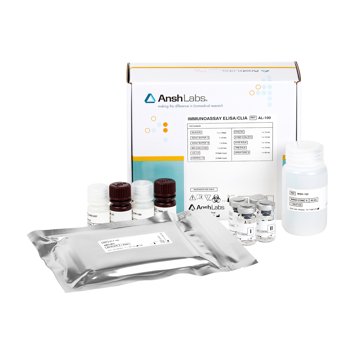
The PCOCheck ELISA is an enzyme-linked immunosorbent assay (ELISA) for the quantitative measurement of anti-Müllerian hormone (AMH) in human serum, Li-Heparin and K2 EDTA plasma. This assay is not impacted by AMH post translational modifications. It is intended to be used as an aid in the determination of ovarian reserve, and stratification of women with polycystic ovarian morphology (PCOM).
| Catalog Numbers | AL-196 (RUO) Instructions For Use |
|---|---|
| Packaging | 96 well microtiter |
| Detection | HRP-based ELISA, colorimetric detection by dual wavelength absorbance at 450 nm and 630 nm as reference filter |
| Dynamic Range | 6, 0.135-24 ng/mL |
| Limit of Detection | 0.029 ng/mL |
| Sample Size | 20 µL |
| Sample Type | Plasma, Serum |
| Species Reactivity | Enquire about animal-specific cross-reactivity |
| Assay Time | 1.5 hours |
| Shelf Life | 24 months |
| Availability | Worldwide |
Anti-Müllerian hormone (AMH), a member of the TGFβ superfamily, is secreted as a 140-kDa homodimeric precursor, consisting of two identical 70-kDa monomers1. Each monomer consists of two parts: (i) a 25-kDa mature C-terminal region, which becomes bioactive after proteolytic cleavage and binds the AMH receptor II (AMHRII), inducing intracellular Smad-signalling2, and (ii) a proregion, which is important in AMH synthesis and the extracellular transport. The AMH precursor is cleaved at amino acid (aa) 451 between the two domains. An additional cleavage site at aa 229 in the pro-region is known, giving rise to three potential cleavage products3. Unlike other members of the TGF-β family, the proregion of the cleaved AMH is critical and enhances the activity of the mature C-terminal when the two cleaved peptides remain associated2-5. AMH undergoes proteolytic cleavage to become biologically active and additional proteolytic processing readily takes place5. This processing, which may differ between individuals, exposes new antigenic sites which may affect measurements as well as AMH epitopes being masked by protein interaction in the circulation5-6. Biological variability can be minimized by the measurement of AMH using liner epitope antibodies that are not impacted by post-translational modification (i.e. glycosylation or proteolytic processing).
AMH is secreted by the Sertoli cells in males. During embryonic development, AMH is responsible for Müllerian duct regression. AMH continues to be produced by the testes until puberty and then decreases slowly to residual post-puberty values. In females, AMH is produced by the granulosa cells of small growing follicles from the 36th week of gestation onwards until menopause when levels become undetectable. The serum concentration of Anti-Müllerian Hormone (AMH) has gained widespread clinical use as a surrogate marker for ovarian reserve7-11. Currently, AMH measurements are used in human fertility counseling7, to predict age of menopause8,9, to diagnose polycystic ovarian syndrome (PCOS)10-12, and to predict response to ovarian stimulation (OS)13,14. Other clinical applications of Anti-Müllerian Hormone (AMH) have been published in ovarian aging15, premature ovarian insufficiency16 ovarian tumors17.18 and many more. As AMH levels may have major implications for clinical decision-making during IVF procedures, egg donation, planning delayed childbearing and attaining optimal ovarian stimulation during treatment, PCOM diagnosis, AMH measurements should be reliable and consistent19.
REFERENCES
1. Pepinski, R.B., et al. (1988) J. Biol. Chem., 263, 18961-18964.
2. di Clemente, N., et al. (2010) Mol Endocrinol, 24 (11): 2193-2206.
3. Donahoe P., (1992) Mol. Reprod. Dev., 32:168–172.
4. Pankhurst MW., et al. (2013) Am J Physiol Endocrinol Metab.; 305: E1241–E1247.
5. Mamsen LS., et al. (2015) Mol Hum Reprod., 21:571–82. doi: 10.1093/molehr/gav024
6. Kawagishi Y., et al. (2017) Mol Reprod Dev., 84:626–37.doi: 10.1002/mrd.22828
7. Seifer DB., et al. (2011) Fertil Steril., 95:747–50.doi: 10.1016/j.fertnstert.2010.10.011
8. Dolleman M., et al. (2013) J Clin Endocrinol Metab., 98:1946–53.doi: 10.1210/jc.201 2-4228
9. Robertson, D.M., et al. (2014) Menopause., 21(12):1277-1286.
10. Iliodromiti S., et al. (2013) J Clin Endocrinol Metab., 98:3332–40.doi: 10.1210/jc.2013-1393
11. Wissing, M.L., et al. (2019) doi: https://doi.org/10.1016/j.fertnstert.2019.03.002
12. Dumont A., et al. (2015) Reprod Biol Endocrinol., 13:137.doi: 10.1186/s12958-015-0134-9
13. Broer SL., et al. (2013) Hum Reprod Update., 19:26–36.doi: 10.1093/humupd/dms041
14. Nelson SM., et al. (2009) Hum Reprod., 24:867–75.doi: 10.1093/humrep/den480
15. Loh JS., et al. (2011) Hum Reprod., 26:2925–32.doi: 10.1093/humrep/der271
16. Méduri G, et. Al.Hum Reprod. 2007; 22:117-23.
17. Lee M, Donahoe., et al. (1996) Journal of Clinical Endocrinology and Metabolism., 81:571-575.
18. Anderson R.A et al. (2006) Human Reproduction., Vol.21 (10): 2583–2592.
19. Antibody compositions and Immunoassay Methods to detect Isoforms of Anti-Mullerian Hormone – AU2013342190
AMH (PCOCheck) ELISA AL-196
Calvert ME, Kalra B, Patel A, Kumar A, Shaw ND. Serum and urine profiles of TGF-β superfamily members in reproductive aged women. Clin Chim Acta. 2022 Jan 1;524:96-100. doi: 10.1016/j.cca.2021.12.002. Epub 2021 Dec 5. PMID: 34875272; PMCID: PMC8740174.
All Products Cited: Inhibin A (pico) ELISA AL-184; Inhibin B ELISA AL-107; Inhibin (Total) ELISA AL-134; AMH (PCOCheck) ELISA AL-196; Activin A ELISA AL-110; Activin B ELISA AL-150; Activin AB ELISA AL-153; Follistatin ELISA AL-117; GDF-9/BMP-15 Complex ELISA AL-181; GDF-15 (Total) ELISA AL-1014; GDF-15 (H-Specific) ELISA AL-1018
Kumar A, Kalra B, Alpadi K, Green K, Wittmaack F. [Audio Presentation] PCOCheck AMH ELISA: A Clinical Case Report to Resolve Mismatched Antral Follicle Counts (AFC) to Serum AMH. Poster presented at Endo SocMeeting; 2021 (Virtual).
All Products Cited: AMH (PCOCHeck)ELISA AL-196
Kumar A, Kalra B, Alpadi K, Green K, Wittmaack F. PCOCheck AMH ELISA: A Clinical Case Report to Resolve Mismatched Antral Follicle Counts (AFC) to Serum AMH. Poster presented at Endo SocMeeting; 2021 (Virtual).
All Products Cited: AMH (PCOCheck) ELISA AL-196
L Melado Vidales, B Lawrenz, A Kumar, B Kalra, B Ata, R Patel, E Kalafat, R Loja Vitorino, I ElKhatib, H Fatemi, O-067 Does the measurement of different AMH isoforms in poor responder patients improve the prediction of ovarian response?, Human Reproduction, Volume 38, Issue Supplement_1, June 2023, dead093.081, https://doi.org/10.1093/humrep/dead093.081
All Products Cited: AMH (PCOCheck) ELISA AL-196; AMH (pico) ELISA AL-124; AMH ELISA AL-105; AMH (Total Mature) ELISA AL-133
