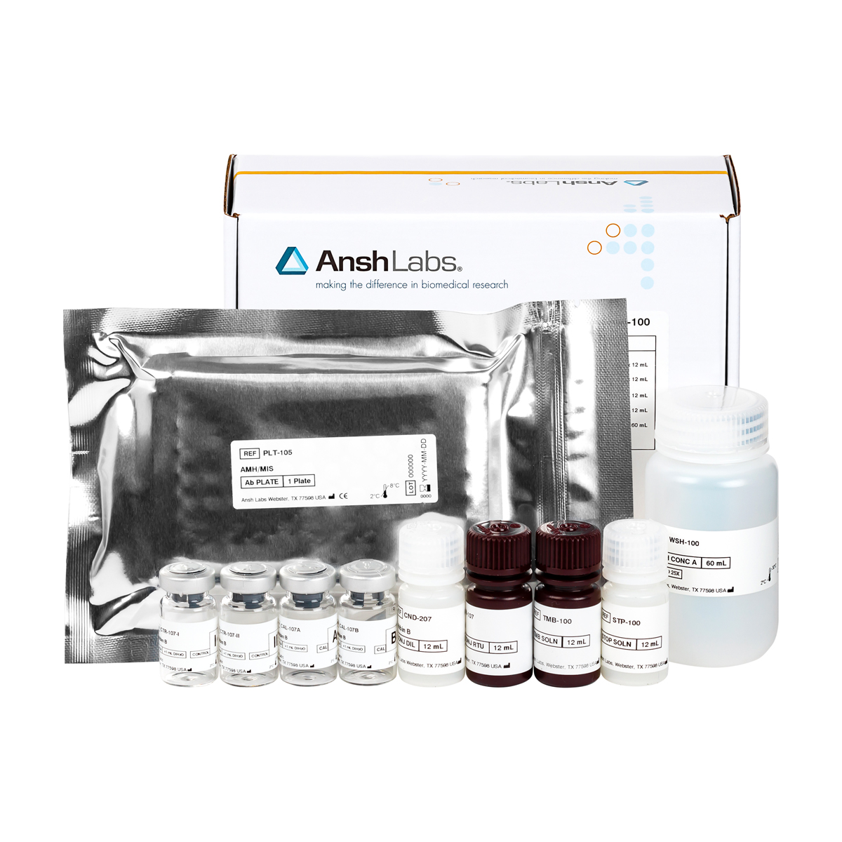
The Unconjugated Estriol Enzyme Immunoassay (EIA) Kit provides materials for the quantitative measurement of unconjugated estriol in serum.
| Catalog Number | AL-138 (FDA) Instructions for Use |
|---|---|
| Packaging | 96 well microtiter |
| Species Reactivity | Enquire about animal-specific cross-reactivity |
| Dynamic Range | 6,0.054-11.75 ng/mL |
| Limit of Detection | 0.04 ng/mL |
| Sample Size | 50 µL |
| Sample Type | Serum |
| Assay Time | 1 hour |
| Availability | Worldwide |
| Shelf Life | 24 months |
| Detection | HRP-based ELISA, colorimetric detection by dual wavelength absorbance at 450 nm and 630 nm as reference filter |
| Storage | Store at 2 to 8°C until expiration date. |
Estriol (1,3,5(10)-estratriene-3,16α,17β-triol; E3) is one of the 3 major naturally occurring estrogens, the others being estradiol and estrone. Estriol is produced almost exclusively during pregnancy, and is the major estrogen produced in the normal human fetus. During pregnancy, the production of estriol depends on an intact maternal-placental-fetal unit.1,2 Steroid precursors from the maternal circulation are taken up by the placenta and converted to progesterone. Progesterone is then converted to dehydroepiandrosterone sulfate (DHEA-S) in the fetal adrenal gland, which is then 16α-hydroxylated in the fetal liver. 16α-hydroxylase activity is present in only very low amounts in placenta and non-fetal tissues. In the placenta, 16α-hydroxy-DHEA-S is then converted sequentially to 16α-hydroxy-DHEA, 16α-hydroxyandrost-4-ene-3,17-dione and, finally, estriol. Estriol may also originate from estrogen precursors, such as 16α-hydroxyestrone. This pathway may account for the high levels of estrone sulfate found in breast cyst fluid.3
Fetal-placental production of estriol leads to a progressive rise in maternal circulating estriol levels, reaching a late-gestational peak which is ~2-3 orders of magnitude greater than non-pregnant levels.1,2 In the maternal circulation, estriol undergoes rapid conjugation in the liver followed by urinary excretion with a half-life of ~20 minutes.1 Therefore, maternal estriol levels can provide a dynamic estimate of fetal production rates. In terms of estrogenic activity, estriol is much less potent than estradiol.2 The physiologic role of estriol is not known.
Specific diagnostic and therapeutic uses for estriol measurements are not completely defined, although clinical utility during pregnancy has been investigated. Since normal estriol production depends on an intact maternal-placental-fetal circulation and functional fetal metabolism, maternal estriol levels have been used to monitor fetal status during pregnancy, particularly during the third trimester. Because estriol concentrations are subject to diurnal and episodic variation, it is common practice to refer serum measurements to a baseline, defined for the patient as either the average or the highest of her three most recent estriol results. However, studies in diabetic pregnancies suggest that low estriol levels have limited value in predicting fetal distress.4 The AL-138 Unconjugated Estriol EIA uses a rabbit anti-estriol antibody preparation with low cross-reactivity to other natural estrogens and estrogen precursors.
References:
1. Buster JE: Gestational changes in steroid hormone biosynthesis, secretion, metabolism, and action. Clin Perinatol 10:527-552, 1983.
2. Cañez MS, Lee KJ, Olive DL: Progestogens and estrogens. Infertil Reproduct Med Clin North Amer 3:59-78, 1992.
3. Levitz M, Raju U, Arcuri F, Brind JL, Vogelman JH, Orentreich N, Granata OM, Castagnetta L: Relationship between the concentrations of estriol sulfate and estrone sulfate in human breast cyst fluid. J Clin Endocrinol Metab 75:726-729, 1992.
4. Miodovnik M, Mimouni F, Hertzberg VS, Siddiqi TA, Tsang RC: Serum unconjugated estriols in insulin-dependent diabetic pregnancies: normative data and clinical relevance. Am J Perinatol 5:327-333, 1988.
5. HHS Publication, 5th ed., 2007. Biosafety in Microbiological and Biomedical Laboratories. Available http://www.cdc.gov/biosafety/publications/bmbl5/BMBL5
6. Approved Guideline – Procedures for the Handling and Processing of Blood Specimens, H18-A3. 2004. Clinical and Laboratory Standards Institute.
7. Kricka L. Interferences in immunoassays – still a threat. Clin Chem 46: 1037–1038, 2000.
