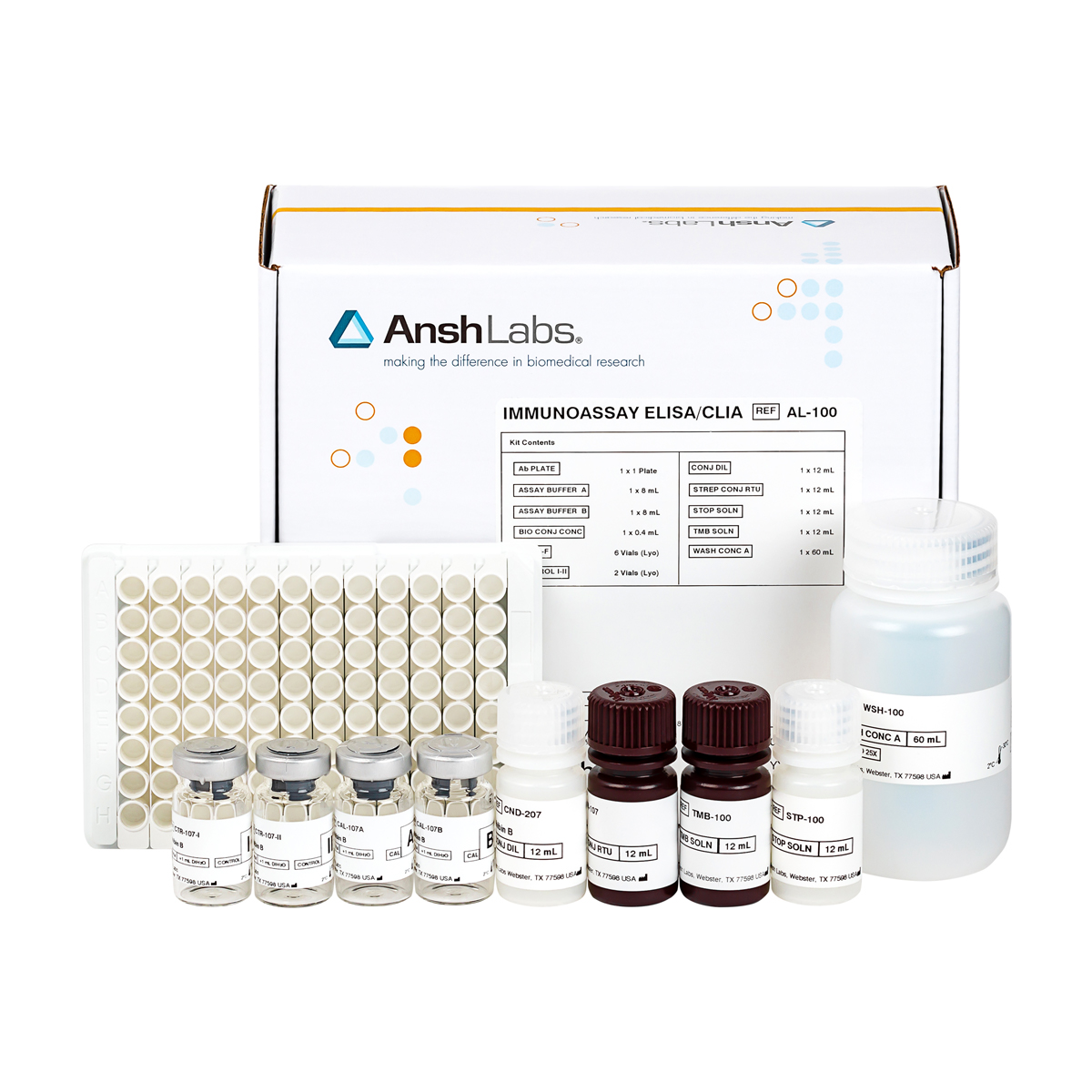
The AFP Enzyme-Linked Immunosorbent Assay (ELISA) Kit provides materials for the quantitative measurement of AFP in serum.
| Catalog Number | |
|---|---|
| Packaging | 96 well microtiter |
| Detection | HRP-based ELISA, colorimetric detection by dual wavelength absorbance at 450 nm and 630 nm as reference filter |
| Dynamic Range | 6, 5-500 ng/mL |
| Limit of Detection | 0.44 ng/mL |
| Species Reactivity | Enquire about animal-specific cross-reactivity |
| Sample Size | 25 uL |
| Sample Type | Serum |
| Assay Time | 1 hour |
| Shelf Life | 24 months |
| Availability | Worldwide |
| Storage | Store at 2 to 8°C until expiration date. |
Alpha-Fetoprotein (AFP) is a 68 kDa protein which is produced primarily during fetal life by the fetal liver yolk sac.1 Elevated AFP levels are seen in patients with nonseminomatous testicular cancer. More than 95% of testicular cancers belong to a heterogeneous group called germ-cell tumors because it is widely believed that they arise in primordial germ cells.2 Germ cell tumors (GCTs) are classified either as seminomatous or as nonseminomatous. The latter can be further classified as embryonal carcinoma, teratoma, or choriocarcinoma. The seminoma histologic subtype can be found in 40% of all germ cell tumors while the nonseminoma histologic subtype can be found in 60% of germ cell tumors.3 The different histologic types of germ cell tumors may occur singly or in various combinations. Elevated AFP levels have been observed in patients diagnosed as having seminomatous testicular cancer with nonseminomatous elements, but not in patients with pure seminoma.4-9
Both AFP and hCG are measured in testicular cancer. Approximately 40% of patients with nonseminomatous germ cell tumors have elevation of only one marker.10 During the clinical course of the disease, the levels of the two markers do not always parallel each other.
A direct relationship has been observed between the incidence of elevated AFP levels in nonseminomatous testicular cancer, and the stage of the disease.4-6 Elevation of AFP (>10 IU/L or 12.1 ng/mL) occurs in 80% of metastatic and in 57% of stage 1 nonseminomatous germ cell tumors.10 In Clinical Stage 2B or higher, AFP and/or hCG are elevated in 65-80% of the cases with increasing frequency according to the bulk of the disease.12
The usefulness of AFP measurements in the management of nonseminomatous testicular cancer patients undergoing cancer therapy has been well established.4,6,13 Current management of testicular germ cell tumors relies upon the use of serum tumor markers, which can indicate the presence of small foci of active tumor that cannot be detected by currently available imaging techniques.10 Markers augment and complement information obtained from radiographic and other staging procedures.14 Also, the short half-lives of tumor markers facilitate their use in assessing tumor burden during therapy. AFP has a serum half-life of 3.5 – 6 days.15 AFP and/or hCG levels are elevated before orchiectomy in about 60% of all Clinical Stage I patients but follow a normal decline after the testicle is removed.12
For patients in clinical remission following treatment, AFP levels generally decrease6. Post-operative AFP levels which fail to return to normal strongly suggest the presence of residual tumor.4,6,16 Following successful resection of primary or metastatic disease, AFP and hCG decline at a rate proportional to their respective half-lives.15 An elevated actual half-life of serum markers following orchiectomy or retroperitoneal lymph node dissection may indicate the presence of occult, persistent disease.14
As recently as the 1970s, nonseminomatous germ cell tumors were often fatal. Due to advances in chemotherapy, most patients are cured, even those with disseminated disease.2 The clinical use of AFP and hCG measurements has been essential to this success. Many patients have a marker surge during the first week of chemotherapy, presumably secondary to tumor lysis. AFP may increase from 20% to 200% over pretreatment levels.14 Chemotherapeutic responses are accompanied by a decline in marker levels. Persistent marker elevation is usually the result of residual malignancy. Rising marker values may occur before or after clinical recurrence and one marker may rise in discordance with the other.14 Tumor recurrence is often accompanied by a rise in serum AFP values prior to clinical evidence of progressive disease.4-5
Elevated serum levels of AFP are also associated with some non-testicular cancers. Increased serum concentrations of AFP were first observed in human subjects with primary heptocellular carcinoma.11 Subsequently, elevated serum AFP values have been associated with other malignant diseases such as teratocarcinoma (with yolk sac components) of the ovary, endodermal sinus tumors, certain gastrointestinal tumors (with and without liver metastasis), and tumors of other tissues.12-13, 16-20 A study performed at the National Institutes of Health and the Mayo Clinic demonstrated elevated AFP values in patients with pancreatic, gastric, colon, and lung cancer.14
In additional studies, AFP was elevated in 60-80% of patients with hepatocellular cancer, in 23% of patients with gastrointestinal cancer and in 10% of patients with liver metastasis from various tumor types.12 However, a normalization of markers does not mean that all viable tumor has been eliminated.14
Notably however, elevated serum AFP concentrations have also been reported in patients with noncancerous diseases such as ataxia telangiectasia, heredity tyrosinemia, neonatal hyperbilirubinemia, acute viral hepatitis, chronic active hepatitis, cirrhosis, and other benign hepatic conditions. 14,16,21-26 AFP is modestly elevated (up to 100 ng/mL) in 20% of patients with non-malignant liver disease.12 Due to its lack of specificity for malignant conditions, AFP testing is not recommended as a screening procedure to detect cancer in the general population.
